Immunohistochemistry and immunofluorescence staining
Table of Contents
Key Summary Table: Immunohistochemistry and Immunofluorescence Staining
| Technique | Principle | Visualization | Advantages | Limitations |
|---|---|---|---|---|
| Immunohistochemistry | Uses antibodies to detect specific proteins | Colorimetric (visible under a light microscope) | Specific, simple, robust | Risk of false positives/negatives |
| Immunofluorescence | Uses antibodies tagged with fluorescent dyes | Fluorescent (requires a fluorescence microscope) | Can visualize multiple proteins simultaneously | Photobleaching, complex interpretation |
Peering into the microscopic world, immunofluorescence staining and immunohistochemistry are like secret decoder glasses, revealing hidden cellular secrets. Stay tuned as we unravel these techniques, making complex science fun and digestible!
Introduction
Ever wondered how scientists visualize the microscopic world? Let’s embark on a journey into the fascinating techniques of immunohistochemistry and immunofluorescence staining. These techniques are like secret keys that unlock the mysteries of the cellular world, revealing the intricate details of cells and tissues that are invisible to the naked eye. They are the tools that medical technologists, like myself, use to understand the complex mechanisms of life and disease.
Immunohistochemistry and immunofluorescence are like two sides of the same coin, each with its unique features.
| Technique | Purpose |
|---|---|
| Immunohistochemistry | Highlights specific cells in a tissue sample |
| Immunofluorescence | Lights up cells in a dazzling display of colors |
What is Immunohistochemistry?
Imagine having a magic lens that can highlight specific cells in a tissue sample. That’s essentially what immunohistochemistry does! It’s a technique that uses antibodies to detect specific proteins in a tissue section. These antibodies are like tiny detectives, each one designed to seek out a specific protein in the cell. When they find their target, they bind to it, marking the spot for us to see under the microscope.
But it’s not just about making cells visible. Immunohistochemistry is also about understanding the function and location of these proteins. By observing where these proteins are located and how they interact with each other, we can gain insights into the inner workings of cells and tissues. This knowledge is crucial in many fields, from research to diagnostics, helping us understand diseases and develop new treatments.

The Method of Immunohistochemistry
| Steps | Description |
|---|---|
| 1. Fixation | Preserves the structure and integrity of the cells and tissues |
| 2. Primary Antibody Application | Antibodies bind to specific proteins |
| 3. Washing | Removes unbound antibodies |
| 4. Secondary Antibody Application | Secondary antibodies bind to primary antibodies |
| 5. Visualization | Areas where antibodies have bound appear as distinct patterns of staining |
The process of immunohistochemistry might seem complex, but let’s break it down into simple steps. First, the tissue sample is prepared and fixed onto a slide. This fixation process is crucial as it preserves the structure and integrity of the cells and tissues. Next, the slide is treated with the specific antibodies. These antibodies are like homing missiles, each designed to seek out and bind to a specific protein.
Once the antibodies have had time to bind to their targets, the slide is washed to remove any unbound antibodies. The next step is the application of a secondary antibody, which is designed to bind to the first antibody. This secondary antibody is tagged with a special marker that can be seen under the microscope. When viewed under the microscope, the areas where the antibodies have bound will appear as distinct patterns of staining, revealing the presence and location of the target protein.
Advantages and Limitations of Immunohistochemistry
Like any scientific method, immunohistochemistry has its strengths and weaknesses. Let’s weigh them up. On the plus side, immunohistochemistry is incredibly specific. Because the antibodies are designed to bind to a specific protein, they can accurately pinpoint the presence and location of that protein in a tissue sample. This specificity makes immunohistochemistry a powerful tool for diagnosing diseases, such as cancer, where the presence or absence of certain proteins can be indicative of the disease.
However, immunohistochemistry is not without its limitations. One of the main challenges is the risk of false positives or negatives. If the antibodies bind to proteins other than their intended target, it can lead to a false positive result. Conversely, if the antibodies fail to bind to their target, it can result in a false negative. Therefore, careful control and validation are crucial in immunohistochemistry to ensure accurate and reliable results.
- Advantages:
- High specificity
- Simple and robust method
- Useful for diagnosing diseases
- Limitations:
- Risk of false positives/negatives
- Requires careful control and validation
What is Immunofluorescence?
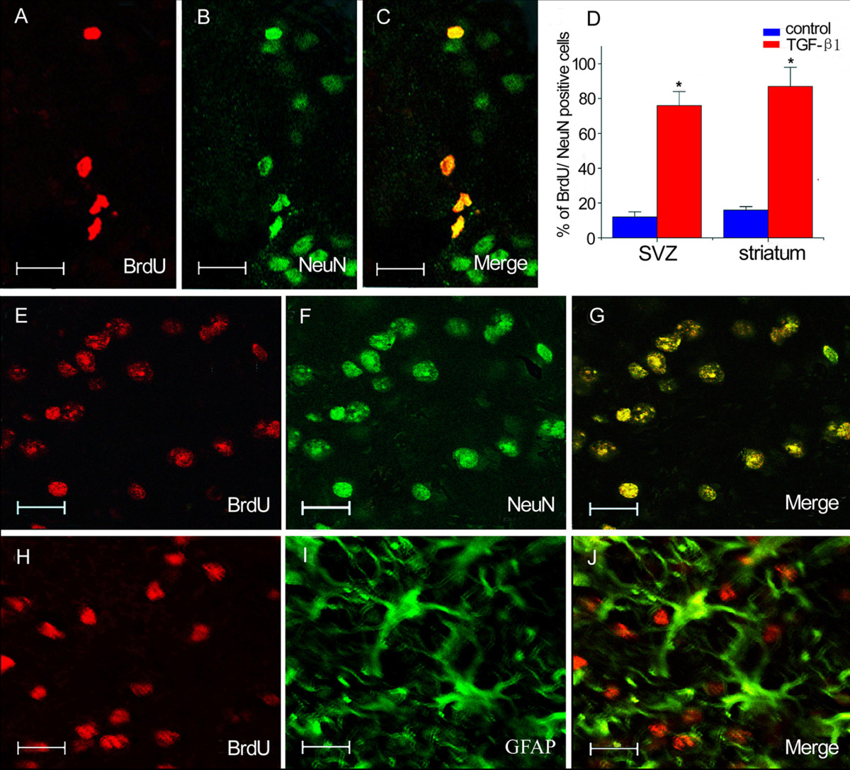
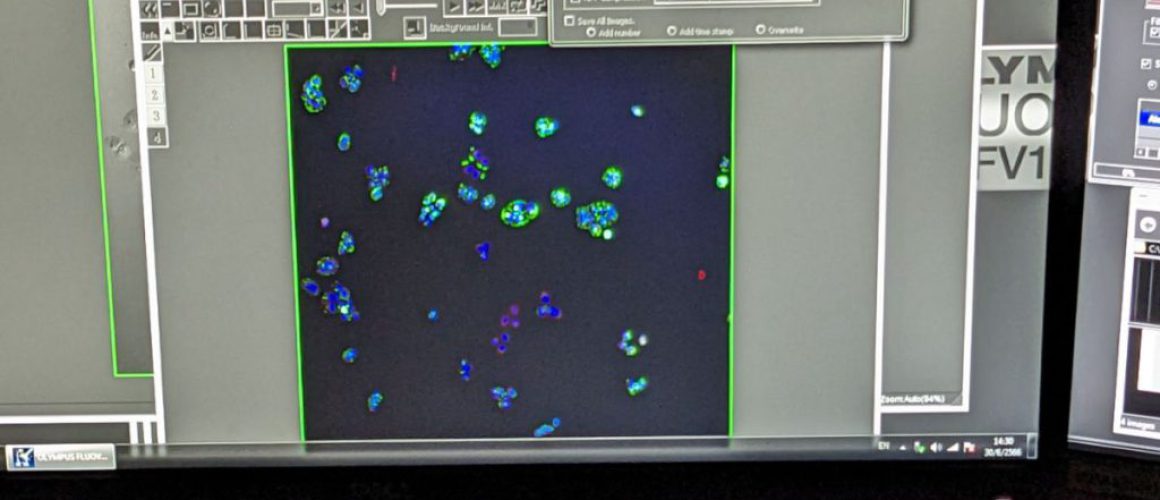
Now, let’s switch gears and explore another powerful technique: immunofluorescence. If immunohistochemistry is like a magic lens, then immunofluorescence is like a spectacular light show. It uses the same principle of using antibodies to detect specific proteins, but in this case, the antibodies are tagged with fluorescent dyes. When exposed to a specific wavelength of light, these dyes emit fluorescence, lighting up the cells in a dazzling display of colors.
Immunofluorescence allows us to see not just the presence of specific proteins, but also their distribution and interaction within the cell. By using different fluorescent dyes, we can label multiple proteins at once, each one lighting up in a different color. This multicolor approach allows us to see how different proteins interact and co-localize within the cell, providing a dynamic view of cellular processes.

The Method of Immunofluorescence
| Steps | Description |
|---|---|
| 1. Fixation | Preserves the structure and integrity of the cells and tissues |
| 2. Antibody Application | Antibodies tagged with fluorescent dyes bind to specific proteins |
| 3. Washing | Removes unbound antibodies |
| 4. Visualization | Fluorescent dyes light up when exposed to specific light wavelength |
Immunofluorescence staining might sound intimidating, but it’s a fascinating process. Let’s simplify it. Much like immunohistochemistry, the process starts with the fixation of the tissue or cells onto a slide. The slide is then treated with the specific antibodies, each tagged with a different fluorescent dye. After a series of washes to remove unbound antibodies, the slide is ready for viewing under a fluorescence microscope.
Under the microscope, the fluorescent dyes light up when exposed to a specific wavelength of light. Each dye emits light at a different wavelength, resulting in different colors. By adjusting the wavelength of light, we can visualize different proteins, each one lighting up in a different color. This colorful display not only makes for stunning images but also provides valuable insights into the cellular processes.
Advantages and Limitations of Immunofluorescence
Immunofluorescence, too, has its pros and cons. Let’s take a closer look. One of the main advantages of immunofluorescence is its ability to visualize multiple proteins simultaneously. By using different fluorescent dyes, we can label and visualize multiple proteins at once, providing a comprehensive view of the cellular landscape.
However, immunofluorescence also has its limitations. One of the main challenges is the photobleaching of the fluorescent dyes. Over time, the dyes can lose their fluorescence when exposed to light, which can affect the quality of the images. Additionally, the interpretation of immunofluorescence images can be complex, requiring expertise and experience to accurately interpret the patterns of fluorescence.
- Advantages:
- Can visualize multiple proteins simultaneously
- Provides a dynamic view of cellular processes
- Limitations:
- Photobleaching of fluorescent dyes
- Requires a fluorescence microscope
- Complex interpretation of images
Comparing Immunohistochemistry and Immunofluorescence
| Aspect | Immunohistochemistry | Immunofluorescence |
|---|---|---|
| Visualization | Colorimetric | Fluorescent |
| Microscope Required | Light microscope | Fluorescence microscope |
| Interpretation | Simpler | More complex |
Now that we’ve explored both techniques, let’s see how they stack up against each other. Both immunohistochemistry and immunofluorescence are powerful techniques for visualizing specific proteins in cells and tissues. They both use the principle of using antibodies to detect specific proteins, but they differ in the way they visualize these proteins.
Immunohistochemistry uses a colorimetric approach, where the antibodies are tagged with enzymes that produce a colored product. This method is simple and robust, and the resulting images can be viewed under a regular light microscope. On the other hand, immunofluorescence uses a fluorescent approach, where the antibodies are tagged with fluorescent dyes. This method provides a more dynamic and detailed view of the cells, but it requires a specialized fluorescence microscope and the images can be more challenging to interpret.
The art of medicine consists in amusing the patient while nature cures the disease.
Voltaire
Conclusion
| Technique | Unique Features |
|---|---|
| Immunohistochemistry | Simple, robust, colorimetric visualization |
| Immunofluorescence | Dynamic, detailed, fluorescent visualization |
Immunohistochemistry and immunofluorescence staining are like two sides of the same coin, each with its unique features. They are powerful tools in the hands of medical technologists, providing valuable insights into the cellular world. Whether it’s diagnosing diseases or unraveling the mysteries of cellular processes, these techniques continue to illuminate our understanding of the microscopic world.
This post is part of my Clinical Microscopy category. Please check out index page on Clinical Microscopy
Other posts of interest: Gram staining and interpretation and Staining in Clinical Microscopy: Revealing the Invisible
Disclaimer: The information provided in this article is for educational purposes only and should not be used as a substitute for professional medical advice.
Frequently Asked Questions
What is immunohistochemistry?
Immunohistochemistry is a technique used in the field of pathology and research that uses antibodies to detect specific proteins in a tissue section. These antibodies are like tiny detectives, each one designed to seek out a specific protein in the cell. When they find their target, they bind to it, marking the spot for us to see under the microscope.
What is immunofluorescence?
Immunofluorescence is another powerful technique that also uses antibodies to detect specific proteins, but in this case, the antibodies are tagged with fluorescent dyes. When exposed to a specific wavelength of light, these dyes emit fluorescence, lighting up the cells in a dazzling display of colors.
What is the difference between immunofluorescence and immunocytochemistry?
While both immunofluorescence and immunocytochemistry use antibodies to detect specific proteins, they differ in the type of samples they analyze. Immunofluorescence is typically used on tissue samples, while immunocytochemistry is used on individual cells.
What is the method of immunohistochemistry?
The method of immunohistochemistry involves several steps, including the preparation and fixation of the tissue sample onto a slide, the application of the specific antibodies, and the visualization of the antibodies under a microscope.
What is the method of immunofluorescence?
The method of immunofluorescence is similar to that of immunohistochemistry. It involves the fixation of the tissue or cells onto a slide, the application of antibodies tagged with fluorescent dyes, and the visualization of the fluorescence under a microscope.
What is the principle of immunohistochemistry?
The principle of immunohistochemistry is based on the specific binding of an antibody to a specific protein in a tissue sample. This specific binding allows for the precise detection and localization of the protein.
What is the principle of immunofluorescence?
The principle of immunofluorescence is similar to that of immunohistochemistry. It also relies on the specific binding of an antibody to a specific protein. However, in immunofluorescence, the antibodies are tagged with fluorescent dyes that emit fluorescence when exposed to a specific wavelength of light.
What are the advantages of immunohistochemistry?
The main advantages of immunohistochemistry are its specificity and simplicity. It allows for the precise detection and localization of specific proteins in a tissue sample, and the method is relatively simple and robust.
What are the advantages of immunofluorescence?
One of the main advantages of immunofluorescence is its ability to visualize multiple proteins simultaneously. By using different fluorescent dyes, we can label and visualize multiple proteins at once, providing a comprehensive view of the cellular landscape.
What are the limitations of immunohistochemistry?
One of the main limitations of immunohistochemistry is the risk of false positives or negatives. If the antibodies bind to proteins other than their intended target, it can lead to a false positive result. Conversely, if the antibodies fail to bind to their target, it can result in a false negative.
What are the limitations of immunofluorescence?
The main limitations of immunofluorescence include the photobleaching of the fluorescent dyes and the complex interpretation of the images. Over time, the dyes can lose their fluorescence when exposed to light, which can affect the quality of the images. Additionally, the interpretation of immunofluorescence images can be complex, requiring expertise and experience.
Further reading
Immunohistochemistry and Immunofluorescence
Sean Schepers is a third-year Medical Technology student at Mahidol University with a passion for all things health and medicine. His journey into the world of medicine has led him to explore various fields. Sean's blog posts offer a unique perspective, combining his academic insights with personal experiences. When he's not studying or blogging, Sean enjoys keeping up with politics and planning his future career in medicine.
In addition to his studies, Sean serves as the chairman of the Rights, Liberties, and Welfare Committee, a role that reflects his commitment to advocacy and social justice. Beyond his academic pursuits, Sean offers tutoring services in English and Biology, further demonstrating his dedication to education and mentorship. His journey is one of continuous discovery, and he invites others to join him as he explores the dynamic and transformative world of medical technology.


