

Histology: A Journey into the Microscopic World
Table of Contents
Introduction
Welcome to the captivating world of histology, where we embark on a remarkable journey into the microscopic realm. Histology, also known as microanatomy, unravels the hidden secrets of our bodies by exploring the intricate structures and functions of tissues and cells. It’s like peering through a powerful microscope, revealing a universe of tiny wonders that make up our being. In this article, we’ll delve into the fascinating realm of histology, its significance in understanding human anatomy, and how it plays a pivotal role in medical diagnostics, research, and education.
In the realm of cells and tissues lies a hidden world waiting to be explored. Histology, the study of microscopic structures, offers a captivating journey into the intricate architecture of the human body.
Unveiling the Mysteries of Tissues
Histology, the captivating science of exploring tissues, takes us on a fascinating journey into the microscopic realm. Tissues serve as the foundation of our organs, orchestrating a symphony of specialized functions to maintain the harmony within our bodies. Under the keen eye of a histologist and the lens of a microscope, the secrets of tissues unfold, revealing intricate structures and cellular arrangements that define their unique identities. From the delicate epithelial linings shielding our organs to the robust muscle fibers propelling us into motion, histology offers us a front-row seat to the mesmerizing tapestry of life.
As we delve into the microscopic world of tissues, we encounter a diverse cast of characters. Epithelial tissues, like a vigilant fortress, form protective barriers and line the surfaces of organs, shielding them from external harm. Connective tissues, with their intricate network of fibers and cells, provide strength, support, and flexibility to the body’s framework. Muscle tissues, ranging from sleek and smooth to formidable and striated, enable graceful movements and vigorous actions. Nervous tissues, like electrical circuits, carry vital messages, orchestrating the symphony of our senses and controlling every aspect of our being. Each tissue type has its own story to tell, revealing its specialized role in the intricate dance of life.
Histology is not merely a study of structures; it offers profound insights into the functions and relationships that drive our physiological processes. Through meticulous observation, histologists unlock the mysteries of tissues, deciphering the interplay between cells, the intricate architecture of blood vessels, and the distribution of specialized cells that maintain our homeostasis. From the pulsating beat of the heart to the rhythmic contraction of muscles, histology unveils the hidden mechanisms that keep us alive and thriving.
The Power of the Microscope: Revealing the Unseen
To truly appreciate the wonders of histology, we need to embrace the power of the microscope. This remarkable tool allows us to magnify and visualize the minute details of tissues, making the invisible visible. By zooming in and panning across the microscopic landscape, we can recognize the intricate architecture of cells, appreciate the diversity of tissues, and understand how their structures correlate with their respective functions. The microscope becomes our portal into a world that would otherwise remain hidden, enabling us to explore the secrets of life at a scale that defies the naked eye.
As we peer through the lens, we witness the extraordinary complexity and organization that underlies every living organism. It is here, in the microscopic realm, that cells engage in a symphony of activity, carrying out their specialized functions and contributing to the overall harmony of the body. We see the elegant branching of nerve cells, the tightly packed arrangement of muscle fibers, and the delicate intricacies of glandular tissues. Each tissue type reveals its unique architecture, reflecting its specific role in maintaining the balance and functionality of the human body.
The microscope not only reveals the structural beauty of tissues but also uncovers the subtle changes that occur in various pathological conditions. It allows us to identify abnormal cellular features, detect signs of disease, and guide medical interventions. By carefully examining tissue samples, histologists and pathologists can unravel the mysteries behind illness, providing invaluable insights that shape diagnoses, treatment plans, and ultimately, patient outcomes. The power of the microscope extends far beyond the boundaries of the laboratory, impacting the lives of countless individuals as it deepens our understanding of human health and disease.
Learning Histology: A Guide to Unlocking the Secrets
Learning histology is akin to embarking on a captivating expedition. To navigate this exciting journey, we need a reliable guide to help us understand the various tissues, their structures, and their functions. A virtual histology guide, accompanied by browser-based tools and interactive visual resources, can serve as an invaluable companion. Through virtual microscopy, we can examine digitized histological specimens, zooming in and out, panning across the slides, and immersing ourselves in the world of histology. This interactive approach enhances our learning experience, allowing us to recognize and appreciate the intricacies of cells and tissues in a dynamic and engaging way.
As we delve into the microscopic world of histology, we encounter a fascinating array of cells and tissues that form the building blocks of the human body. With each slide we examine, a new world unfolds, revealing the intricate beauty and complexity hidden within. We learn to decipher the unique structures and functions of epithelial tissues, connective tissues, muscle tissues, and nervous tissues. Our guide takes us on a tour of the microscopic landscapes, introducing us to the specialized cells that enable our organs to function harmoniously.
Through this journey, we realize the significance of histology in our understanding of diseases and their diagnosis. We witness the profound impact that cellular changes can have on our health, as histological analysis helps identify abnormalities, inflammation, and even the presence of cancerous cells. Armed with this knowledge, we gain a deeper appreciation for the role of histologists and pathologists in unraveling the mysteries behind various medical conditions.
Comparison of Different Histological Techniques
| Technique | Description |
|---|---|
| Hematoxylin and Eosin Staining | The most widely used staining method in histology, highlighting nuclei (blue) and cytoplasm (pink) |
| Periodic Acid-Schiff Staining | Stains carbohydrates pink or magenta, often used to identify glycogen-rich structures |
| Masson’s Trichrome Staining | Distinguishes collagen fibers (blue/green), muscle fibers (red), and cell nuclei (dark brown/black) |
| Immunohistochemistry | Utilizes antibodies to detect specific proteins, aiding in the identification of cell types and biomarkers |
| In Situ Hybridization | Visualizes specific RNA molecules within cells, providing insights into gene expression patterns |
Exploring the Histology Laboratory: Where Science Comes Alive
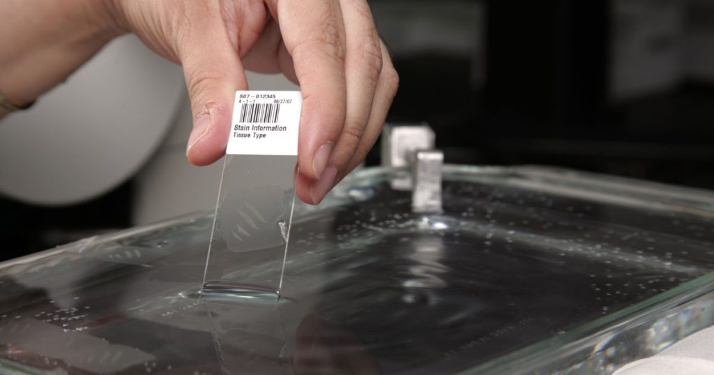
No exploration of histology is complete without a glimpse into the histology laboratory, where science comes alive. As I step into this controlled environment, I am immediately struck by the sense of purpose and dedication that permeates the air. The laboratory is a bustling hub of activity, with skilled histotechnologists meticulously working their magic on tissue samples, transforming them into delicate slides ready for microscopic examination.
Each step in the histology process is carried out with meticulous precision. From the moment a tissue specimen arrives, it undergoes a series of carefully orchestrated procedures. First, the specimen is meticulously fixed to preserve its delicate structure and prevent decay. Then, it is carefully embedded in paraffin or other embedding materials, ensuring that it is solidified and can be sliced into thin sections without distortion.
Sectioning is a true art form, requiring steady hands and sharp instruments. Histotechnologists skillfully cut ultra-thin slices of tissue, each no thicker than a whisper, using microtomes. These delicate sections are then meticulously mounted on glass slides, ready to be transformed into visual masterpieces that unlock the mysteries of microanatomy.
The histology laboratory is a testament to the dedication and expertise of these professionals. Their unwavering commitment to detail and quality is evident in every slide they produce. It is here, in this realm of precision and technique, that the invisible becomes visible, and the hidden intricacies of tissues and cells are revealed. The histology laboratory is where science truly comes alive, illuminating the wonders of the microscopic world and paving the way for breakthroughs in medical research, diagnostics, and our understanding of the human body.
Histology: A Gateway to Medical Diagnosis and Research
Histology plays a crucial role in medical diagnostics, offering insights into the nature of diseases and aiding in accurate diagnosis. By examining abnormal tissue samples, histopathologists can identify cellular and structural changes associated with various conditions. From cancerous growths to inflammatory processes, histology allows us to visualize the effects of diseases at the cellular level, providing essential information for treatment planning and patient care. Moreover, histological research contributes to groundbreaking discoveries, deepening our understanding of diseases, and driving advancements in medical science.
As a medical technology student, I am fascinated by the intricate world that histology unveils. Peering through a microscope, I witness the hidden beauty and complexity of tissues and cells. Histology acts as a gateway, allowing us to explore the microcosm within our bodies and unravel the mysteries that lie beneath the surface. It is through histological analysis that diseases reveal their distinctive features, allowing healthcare professionals to make informed decisions and tailor treatment strategies accordingly.
The significance of histology extends beyond diagnosis. It fuels medical research, serving as a foundation for breakthroughs and innovations. By studying tissue samples, researchers gain valuable insights into disease mechanisms, test new therapeutic approaches, and explore the potential of regenerative medicine. Histology contributes to the development of cutting-edge techniques and technologies that shape the future of healthcare.
Essential Skills for Histologists
- Microscope Handling and Operation
- Slide Preparation Techniques
- Staining and Mounting Methods
- Microscopic Observation and Analysis
- Knowledge of Cell and Tissue Structures
- Attention to Detail and Accuracy
- Proficiency in Digital Imaging and Analysis Software
- Understanding of Histological Artifacts and Troubleshooting
- Familiarity with Laboratory Safety and Protocols
- Continuous Learning and Adaptability to New Techniques
Call to Action
Embark on your own journey into the microscopic world of histology. Explore the captivating beauty and complexity of tissues and cells, and unravel the secrets they hold. Visit seanschepers.com/histology for more resources, articles, and insights into this captivating field of study. Let the wonders of histology ignite your curiosity and deepen your understanding of the intricate mechanisms that sustain life.
Histology is like reading a captivating story where cells and tissues come alive, revealing the intricate details that make us who we are.
Conclusion
Histology invites us to peer through the microscope and embark on a journey into the microscopic world. By understanding the structures and functions of tissues and cells, we gain a profound appreciation for the remarkable complexity and harmony that exists within our bodies. Histology empowers medical professionals, researchers, and students to unlock the mysteries of diseases, make accurate diagnoses, and drive advancements in healthcare. So, let’s embrace this journey into the microscopic world, where every slide reveals a new story waiting to be explored.
Disclaimer: The above article provides an overview of histology and its significance, but please consult with qualified professionals or experts for any specific medical advice or concerns.
Frequently Asked Questions
What is histology?
Histology, also known as microanatomy, is the study of microscopic structures, including cells and tissues, using specialized techniques and tools such as microscopes. It provides insights into the organization, structure, and function of different components of the human body.
Why is histology important in the field of medicine?
Histology plays a vital role in medicine as it helps in understanding the normal structure and functioning of tissues and organs. It also enables the identification of abnormal cellular changes, aiding in the diagnosis and treatment of various diseases and conditions.
How is histology different from pathology?
Histology focuses on the study of normal tissue structures and their functions, while pathology examines changes or abnormalities within tissues to diagnose diseases. Pathologists often use histological techniques to analyze tissue samples and provide insights into the nature and progression of diseases.
What are some common staining techniques used in histology?
Staining techniques are essential in histology to enhance the visualization of tissue components. Hematoxylin and eosin (H&E) staining, Periodic Acid-Schiff (PAS) staining, and Masson’s trichrome staining are commonly employed to highlight different cellular structures and substances within tissues.
Can histology be studied virtually?
Yes, with the advancement of technology, virtual microscopy has emerged as a valuable tool for studying histology. Online platforms and virtual microscopy software allow learners to explore digitized histological slides, providing an interactive and immersive learning experience.
What career opportunities are available in histology?
Histology offers various career paths, including histotechnologists, histology technicians, and histopathologists. These professionals work in medical laboratories, research institutions, hospitals, and academic settings, performing tasks related to tissue processing, slide preparation, and histological analysis.
How does histology contribute to medical research?
Histology plays a crucial role in medical research by providing insights into disease mechanisms, evaluating treatment responses, and studying the effects of new therapies. It helps researchers identify cellular changes, biomarkers, and tissue interactions that can lead to advancements in diagnostics and therapeutics.
Can histological findings change medical treatment decisions?
Absolutely. Histological findings can provide critical information that influences medical treatment decisions. By analyzing tissue samples, histopathologists can identify specific cellular characteristics, disease progression, or the presence of malignancy, guiding healthcare professionals in selecting appropriate treatment options for patients.
Sources
Histology Guide – virtual microscopy laboratory

Sean Schepers is a third-year Medical Technology student at Mahidol University with a passion for all things health and medicine. His journey into the world of medicine has led him to explore various fields. Sean's blog posts offer a unique perspective, combining his academic insights with personal experiences. When he's not studying or blogging, Sean enjoys keeping up with politics and planning his future career in medicine.
In addition to his studies, Sean serves as the chairman of the Rights, Liberties, and Welfare Committee, a role that reflects his commitment to advocacy and social justice. Beyond his academic pursuits, Sean offers tutoring services in English and Biology, further demonstrating his dedication to education and mentorship. His journey is one of continuous discovery, and he invites others to join him as he explores the dynamic and transformative world of medical technology.