

Gram staining and interpretation
Table of Contents
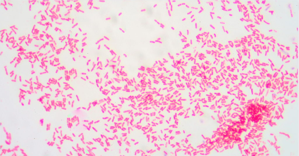
Gram Staining
| Property | Gram-Positive Bacteria | Gram-Negative Bacteria |
|---|---|---|
| Cell Wall Composition | Thick peptidoglycan layer | Thin peptidoglycan layer and an outer membrane |
| Gram Stain Retention | Retains crystal violet dye, appears purple | Does not retain crystal violet dye, takes up safranin counterstain, appears red/pink |
| Antibiotic Susceptibility | Generally more susceptible to antibiotics like penicillin | Generally more resistant due to outer membrane |
Ever played with color-changing squishy toys? Gram staining in medical technology is a bit like that, but with bacteria! It’s a fascinating process that helps us categorize these microscopic organisms. Stick around to unravel the mystery of this color-coded world!

Introduction
When it comes to identifying bacteria, one of the most fundamental techniques used in clinical microscopy is Gram staining. This method, named after the Danish bacteriologist Hans Christian Gram, is a cornerstone in the field of bacteriology. It provides critical information about the bacterial cell wall’s physical properties, which can be a decisive factor in the treatment of bacterial infections. So, what exactly is Gram staining, and what does it indicate? Let’s explore.
Advantages of Gram Staining
- Quick and easy to perform.
- Provides preliminary information about the nature of a bacterial infection.
- Guides the selection of appropriate antibiotics for treatment.
- Helps in the identification of the bacteria species.
What is Gram Staining?
Imagine you’re sorting a bag of mixed candies into their respective types. Gram staining works similarly, but instead of candies, we’re dealing with bacteria. This technique, named after Hans Christian Gram, sorts bacteria into two major groups: gram-positive and gram-negative. It’s like a secret decoder, revealing the structure of their cell walls. The key player here is peptidoglycan, a substance found in varying amounts in different bacteria. Gram-positive bacteria have a thick peptidoglycan layer, while gram-negative ones have a thinner layer.
Now, let’s talk about the star of the show, the crystal violet dye. This dye is like a spotlight that shines on the bacteria, but only some can hold onto it. Gram-positive bacteria, with their thick peptidoglycan layer, hold onto the dye and turn purple. On the other hand, gram-negative bacteria, with their thinner layer, can’t hold onto the dye and remain colorless… for now.
Differences Between Gram-Positive and Gram-Negative Bacteria
| Property | Gram-Positive Bacteria | Gram-Negative Bacteria |
|---|---|---|
| Cell Wall Thickness | Thick | Thin |
| Outer Membrane | Absent | Present |
| Teichoic Acids | Present | Absent |
| Lipopolysaccharides | Absent | Present |
| Periplasmic Space | Absent | Present |
The Process of Gram Staining
| Step | Description |
|---|---|
| 1. Crystal Violet | The primary stain that colors all cells purple |
| 2. Gram’s Iodine | The mordant that forms a complex with crystal violet, getting trapped in the cell wall |
| 3. Decolorization | Alcohol or acetone is used to wash away the dye. Gram-positive cells remain purple, Gram-negative cells lose the color |
| 4. Safranin | The counterstain that is absorbed by Gram-negative cells, making them appear red/pink |
| 5. Microscopic Examination | Determine the color of the cells to classify them as Gram-positive or Gram-negative |
The process of Gram staining involves several steps that result in bacteria being stained either purple (gram-positive) or pink/red (gram-negative). The first step is the application of a purple dye, known as crystal violet, to a heat-fixed smear of bacterial culture. Next, a mordant, Gram’s iodine, is added to form a complex between the crystal violet and iodine. This complex gets trapped within the multilayered peptidoglycan meshwork of gram-positive bacteria.
The third step is the decolorization step, which is the most critical step as it differentiates between gram-positive and gram-negative bacteria. During decolorization, a solution of alcohol or acetone is added. If the thick peptidoglycan layer in gram-positive bacteria retains the crystal violet dye, it remains purple. In contrast, the thinner peptidoglycan layer of gram-negative bacteria does not retain the dye and is decolorized.
The final step is the application of a counterstain, usually safranin, a red dye. This dye is not absorbed by gram-positive bacteria, but it is absorbed by gram-negative bacteria, making them appear red or pink under a microscope.

The Importance of Gram Staining in Medical Technology
Think of Gram staining as the first line of defense in the battle against bacterial infections. It’s like a detective, providing us with the first clues about the identity of the bacteria causing the infection. This information is crucial because different types of bacteria respond to different antibiotics. For example, gram-positive bacteria are usually more susceptible to antibiotics like penicillin, while gram-negative bacteria, with their protective outer membrane, are often more resistant.
But the Gram stain doesn’t work alone. It’s part of a team, working alongside other tests that look at the bacteria’s shape and oxygen requirements. Together, they help us identify the exact species of bacteria we’re dealing with. In the world of medical technology, where accurate and timely identification of pathogens can mean the difference between life and death, the Gram stain is an invaluable tool.
Common Diseases Caused by Gram-Positive and Gram-Negative Bacteria
| Gram-Positive Bacteria | Diseases | Gram-Negative Bacteria | Diseases |
|---|---|---|---|
| Staphylococcus aureus | Skin infections, pneumonia | Escherichia coli | Urinary tract infections, food poisoning |
| Streptococcus pneumoniae | Pneumonia, meningitis | Salmonella typhi | Typhoid fever |
| Bacillus anthracis | Anthrax | Pseudomonas aeruginosa | Wound and burn infections |
| Clostridium tetani | Tetanus | Neisseria gonorrhoeae | Gonorrhea |
Factors Affecting the Results of Gram Staining
- Quality of the reagents used.
- Age and condition of the bacterial culture.
- Correct execution of the decolorization step.
- Quality of the microscope used for examination.
Gram Staining: Interpretation and Challenges
Interpreting Gram stain results is a bit like solving a puzzle. The pieces are the color changes we observe. Gram-positive bacteria hold onto the crystal violet dye and appear purple, while Gram-negative bacteria take up the counterstain safranin and appear red or pink. But like any good puzzle, there are challenges.
Not all bacteria play by the rules. Some don’t stain well with the Gram stain and are called Gram-variable or Gram-indeterminate. It’s like they’ve hidden some of their puzzle pieces. Also, the age of the bacterial culture can throw a wrench in the works. Older cultures of some Gram-positive bacteria may not hold onto the crystal violet stain as well and might appear Gram-negative. So, while the Gram stain is a powerful tool, it’s not without its quirks and challenges.
Limitations of Gram Staining
- Not all bacteria can be definitively classified by this technique.
- Some bacteria do not stain well with the Gram stain and are termed Gram-variable or Gram-indeterminate.
- The age of the bacterial culture can affect the results of the Gram stain.
- Requires a fresh culture of bacteria for accurate results.
Conclusion
While it may seem like a simple coloring process, Gram staining is a powerful tool in clinical microscopy that has significant implications for diagnosing and treating bacterial infections. It’s a testament to the ingenuity of scientists like Hans Christian Gram, who, with a simple staining technique, provided us with a method to visualize and identify the microscopic enemies that we battle in the field of healthcare.
This post is part of my Clinical Microscopy category. Please check out index page on Clinical Microscopy
Other pages of interest: Fungal staining (KOH, Calcofluor White) and interpretation and Immunohistochemistry and immunofluorescence staining
This post is part of my Bacteriology category. Also check out my index page on Bacteriology.
Other pages of interest: Acid-fast staining (Ziehl-Neelsen, Kinyoun) and interpretation and The Role of Bacteriology in Disease Diagnosis
Disclaimer: This article is intended for informational purposes only and is not a substitute for professional medical advice or consultation. Always seek the advice of your healthcare provider with any questions you may have regarding a medical condition or treatment.
Frequently Asked Questions
What is the basic principle of Gram staining?
The basic principle of Gram staining is the differential staining of bacteria based on the properties of their cell walls. Gram-positive bacteria have a thick layer of peptidoglycan in their cell walls that retains the crystal violet dye used in the staining process, while Gram-negative bacteria have a thinner peptidoglycan layer and do not retain this dye.
How is Gram staining used to identify bacteria?
Gram staining is used to classify bacteria into two major groups: Gram-positive and Gram-negative. This classification can provide preliminary information about the nature of a bacterial infection, guiding the selection of appropriate antibiotics for treatment. It also helps in the identification of the bacteria species when combined with other information such as the bacteria’s shape and oxygen requirements.
How can you tell the difference between Gram-positive and Gram-negative bacteria?
Gram-positive bacteria retain the crystal violet dye used in the Gram staining process and appear purple under a microscope. On the other hand, Gram-negative bacteria do not retain this dye during the decolorization step and take up the red counterstain safranin, causing them to appear red or pink under a microscope.
What is the conclusion of the Gram stain?
The conclusion of the Gram stain is the determination of whether the bacteria are Gram-positive (purple) or Gram-negative (red or pink). This information is crucial in the field of healthcare for diagnosing and treating bacterial infections.
What are the 5 steps of gram staining?
The five steps of Gram staining are:
1. Application of the primary stain, crystal violet.
2. Addition of the mordant, Gram’s iodine, which forms a complex with the crystal violet that gets trapped within the cell wall.
3. Decolorization with alcohol or acetone, which removes the crystal violet-iodine complex from Gram-negative bacteria but not from Gram-positive bacteria.
4. Application of the counterstain, safranin, which is not absorbed by Gram-positive bacteria but is absorbed by Gram-negative bacteria, making them appear red or pink.
5. Examination under a microscope to determine the color of the bacteria and hence their Gram reaction.
Why do gram negative bacteria appear pink after gram staining?
Gram-negative bacteria appear pink after Gram staining because they do not retain the primary crystal violet dye during the decolorization step due to their thinner peptidoglycan layer and the presence of an outer membrane. Instead, they take up the counterstain, safranin, which gives them a pink or red color when viewed under a microscope.
What is Gram staining?
Gram staining is a laboratory method used to classify bacteria into two major groups: gram-positive and gram-negative. This classification is based on the chemical and physical properties of their cell walls, primarily the ability to retain a violet dye called crystal violet.
Who invented the Gram staining technique?
The Gram staining technique was invented by a Danish bacteriologist named Hans Christian Gram in 1884. It’s named after him in recognition of his significant contribution to bacteriology.
How does Gram staining work?
Gram staining works by applying a series of dyes that cause gram-positive bacteria (those with a thick peptidoglycan cell wall) to turn purple and gram-negative bacteria (those with a thin peptidoglycan cell wall) to turn pink or red. The process involves four steps: staining with crystal violet, adding a mordant (Gram’s iodine), decolorization with alcohol or acetone, and counterstaining with safranin.
What is the difference between Gram-positive and Gram-negative bacteria?
The difference between Gram-positive and Gram-negative bacteria lies in the structure of their cell walls. Gram-positive bacteria have a thick peptidoglycan layer that retains the crystal violet dye, making them appear purple under a microscope. On the other hand, Gram-negative bacteria have a thinner peptidoglycan layer and an outer membrane, causing them to lose the crystal violet dye during decolorization and take up the red counterstain safranin.
Why is Gram staining important in medical technology?
Gram staining is a crucial tool in medical technology as it provides preliminary information about the nature of a bacterial infection. This information can guide the selection of appropriate antibiotics for treatment. It also helps in the identification of the bacteria species when combined with other information such as the bacteria’s shape and oxygen requirements.
Are there any limitations to Gram staining?
Yes, there are limitations. Not all bacteria can be definitively classified by this technique. Some bacteria do not stain well with the Gram stain and are termed Gram-variable or Gram-indeterminate. Moreover, the age of the bacterial culture can affect the results of the Gram stain. Older cultures of some Gram-positive bacteria may not retain the crystal violet stain well and may appear Gram-negative.
Further reading
Gram Staining: Principle, Procedure, Interpretation, …

Sean Schepers is a third-year Medical Technology student at Mahidol University with a passion for all things health and medicine. His journey into the world of medicine has led him to explore various fields. Sean's blog posts offer a unique perspective, combining his academic insights with personal experiences. When he's not studying or blogging, Sean enjoys keeping up with politics and planning his future career in medicine.
In addition to his studies, Sean serves as the chairman of the Rights, Liberties, and Welfare Committee, a role that reflects his commitment to advocacy and social justice. Beyond his academic pursuits, Sean offers tutoring services in English and Biology, further demonstrating his dedication to education and mentorship. His journey is one of continuous discovery, and he invites others to join him as he explores the dynamic and transformative world of medical technology.