
")
Fungal staining (KOH, Calcofluor White) and interpretation
Table of Contents
Key Summary Table: Fungal Staining
| Staining Technique | Purpose | Key Ingredient | Advantage |
|---|---|---|---|
| Calcofluor White Stain | Detecting fungi in clinical samples | Calcofluor White | Fluoresces under UV light, enhancing visibility of fungi |
| KOH Stain | Clearing non-fungal elements from a sample | Potassium Hydroxide (KOH) | Enhances visibility and identification of fungi |
Fungal staining, specifically with KOH and Calcofluor White, is like a secret decoder ring for the microscopic world of fungi. Stay tuned as we unravel the mysteries of these staining techniques and their interpretations, all explained in layman’s terms!
Introduction
Welcome to the captivating world of fungal staining, a cornerstone in the realm of clinical microscopy. This technique, as intriguing as it is essential, allows us to visualize and identify fungi, those microscopic organisms that play such a significant role in our world. From the fungi that contribute to the decomposition process in nature to those that cause infections in humans, understanding these organisms is crucial. And it all starts with the ability to see them clearly.
The world of fungi is a microscopic universe, full of unseen wonders. And it’s through techniques like fungal staining that we get a glimpse into this world.
Fungal staining is not just about making the invisible visible. It’s about bringing to light the intricate details of these organisms, their structure, their form, and their unique characteristics. It’s about transforming a seemingly ordinary sample into a vibrant, detailed image that tells a story. A story of life at a microscopic level, a story that could lead to a better understanding of diseases, and ultimately, to better health outcomes.
In this article, we’ll journey through the world of fungal staining, exploring its techniques, its purpose, and its significance. We’ll delve into the specifics of two popular methods – KOH and Calcofluor White – and discuss their interpretation. So, let’s get started on this fascinating journey.
The Basics of Fungal Staining
| Fungal Staining Technique | Commonly Used For |
|---|---|
| Calcofluor White Stain | Detecting fungi in clinical samples |
| KOH Stain | Clearing non-fungal elements from a sample |
Fungal staining is a specialized technique that brings the world of fungi to life. It’s like a magic wand that reveals the hidden details of these microscopic organisms. But how does it work? And why is it so important?
Fungal staining works by using specific dyes that bind to the components of the fungal cell. This binding makes the fungi visible under a microscope, allowing us to see their structure and form. But it’s not just about visibility. The way these dyes bind can also tell us a lot about the type of fungi we’re dealing with, aiding in their identification.
The importance of fungal staining cannot be overstated. In the field of medical technology, it’s a crucial tool for diagnosing fungal infections. By identifying the type of fungi present in a sample, healthcare professionals can determine the most effective treatment strategy.
- Fungal staining techniques are crucial for visualizing and identifying fungi.
- Different staining techniques provide different insights about the fungi.
- The choice of staining technique depends on the type of fungi and the specific information needed.
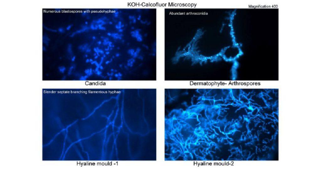
Calcofluor White Stain: A Powerful Tool for Detecting Fungi
| Property | Description |
|---|---|
| Fluorescence | Glows under UV light |
| Binding | Binds to chitin and cellulose in fungal cell walls |
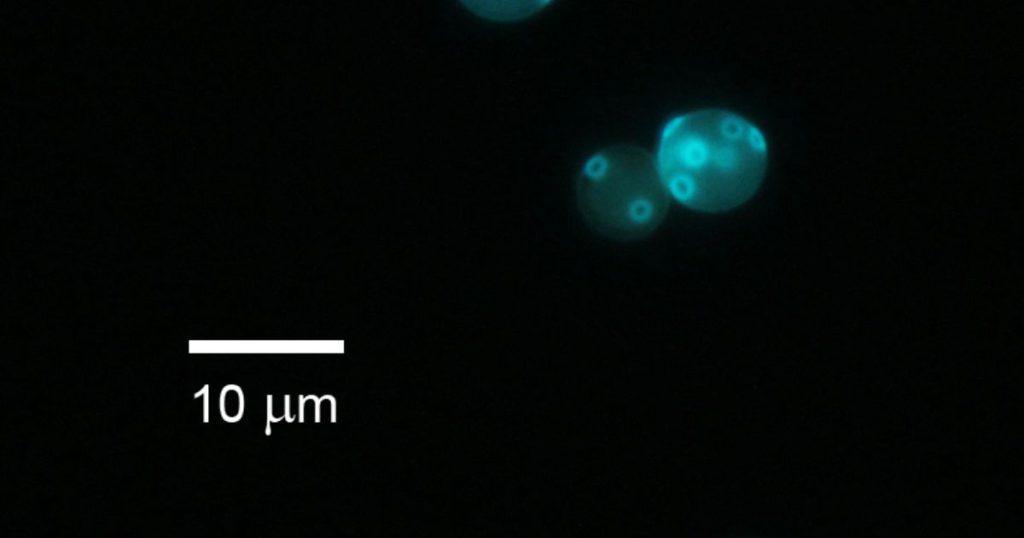
Among the various staining techniques, Calcofluor White stain holds a special place. This fluorescent dye has a unique ability to bind to certain types of fungi, illuminating them under a microscope.
Calcofluor White works by binding to the chitin and cellulose in the cell walls of fungi. When exposed to ultraviolet light, the dye fluoresces, making the fungi stand out against the background. This makes it easier to identify the presence of fungi in a sample, even when they are present in small numbers.
But the power of Calcofluor White stain goes beyond just detection. It also provides valuable information about the structure of the fungi, contributing to their identification. This makes it a powerful tool in the diagnosis of fungal infections, helping to guide treatment decisions.
- Calcofluor White stain binds to chitin and cellulose in fungal cell walls.
- The stain fluoresces under UV light, making the fungi stand out.
- The binding patterns of the stain can provide insights into the type of fungi.
The Purpose of Adding Calcofluor White to a Fungal Preparation
Adding Calcofluor White to a fungal preparation is like adding a high-powered spotlight to a stage. It illuminates the fungi, making them the stars of the show. But there’s more to this process than just making things brighter.
When added to a fungal preparation, Calcofluor White binds to the chitin and cellulose in the fungal cell walls. This binding is not random. It occurs in specific patterns that reflect the structure of the fungi. By observing these patterns under a microscope, we can gain insights into the type of fungi we’re dealing with.
Moreover, the use of Calcofluor White enhances the contrast between the fungi and the background. This makes it easier to identify the presence of fungi, even when they are present in small numbers. This is particularly important in clinical settings, where early detection of fungal infections can make a significant difference in patient outcomes.
KOH in Fungal Identification: A Key Ingredient
| Property | Description |
|---|---|
| Base | Strong base that dissolves non-fungal elements |
| Enhancement | Enhances staining process, making fungi more visible |
Potassium hydroxide, or KOH, might seem like an odd ingredient in the world of fungal identification. After all, it’s a strong base, more commonly associated with soap making or drain cleaning. But in the realm of fungal staining, KOH plays a crucial role.
KOH is used in fungal staining to dissolve the non-fungal elements in a sample. This leaves behind the fungal cells, making them easier to see under a microscope. But KOH does more than just clear the stage for the fungi. It also enhances the staining process, making the fungi more visible and easier to identify.
The use of KOH in fungal identification is a testament to the power of chemistry in the field of medical technology. It’s a reminder that sometimes, the most effective tools are the ones that seem the least likely.
- KOH is a strong base that dissolves non-fungal elements in a sample.
- The use of KOH makes the fungal cells easier to see under a microscope.
- KOH enhances the staining process, making the fungi more visible and easier to identify.
The Best Stain for Fungal Elements: A Comparative Study
| Staining Technique | Advantage |
|---|---|
| Calcofluor White Stain | Fluoresces under UV light, enhancing visibility of fungi |
| KOH Stain | Clears non-fungal elements, enhancing visibility of fungi |
With so many staining techniques available, which one is the best for visualizing fungal elements? The answer to this question is not straightforward. It depends on the type of fungi, the nature of the sample, and the specific information needed.
For example, Calcofluor White stain is excellent for detecting fungi in clinical samples, thanks to its ability to fluoresce under ultraviolet light. On the other hand, KOH is particularly useful for clearing non-fungal elements from a sample, making the fungi easier to see.
In some cases, a combination of staining techniques may be used. This can provide a more comprehensive view of the fungi, aiding in their identification. The choice of staining technique is a critical decision in the process of fungal identification, one that requires a deep understanding of the strengths and limitations of each method.
- Different staining techniques have different strengths and limitations.
- The choice of staining technique depends on the type of fungi and the nature of the sample.
- A combination of staining techniques may provide a more comprehensive view of the fungi.
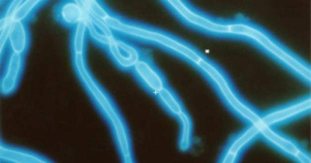
The Principle of Calcofluor Staining: A Closer Look
The principle of Calcofluor staining is a fascinating blend of chemistry and biology. It’s a process that transforms a seemingly ordinary sample into a vibrant, detailed image that tells a story.
Calcofluor White is a fluorescent dye that binds to the chitin and cellulose in the cell walls of fungi. When exposed to ultraviolet light, the dye fluoresces, illuminating the fungi. This makes them easier to see under a microscope, aiding in their identification.
But the principle of Calcofluor staining goes beyond just making things brighter. The way the dye binds to the fungi provides valuable information about their structure. This can help in distinguishing between different types of fungi, contributing to their identification.
KOH and Tinea Infection: A Diagnostic Duo
KOH is often used in the lab diagnosis of tinea infection, a common fungal infection that affects the skin. But why is KOH such a valuable tool in this context?
Tinea infections are caused by a group of fungi known as dermatophytes. These fungi have a unique characteristic: they produce an enzyme that allows them to digest keratin, a protein found in the skin, hair, and nails. This makes the skin a perfect feeding ground for these fungi, leading to infection.
KOH, with its ability to dissolve non-fungal elements, is particularly effective in visualizing dermatophytes in skin samples. By clearing away the keratin and other debris, KOH allows the fungal cells to stand out, making them easier to identify under a microscope.
The power of science is not just in understanding the world around us, but also in developing tools that allow us to see the unseen.
Conclusion
Fungal staining techniques, such as KOH and Calcofluor White, are powerful tools in the field of clinical microscopy. They allow us to visualize and identify fungi, contributing to the diagnosis and treatment of fungal infections.
But these techniques are more than just tools. They are a testament to the power of science and technology in improving healthcare outcomes. They remind us that even the smallest organisms, like fungi, have a significant impact on our health and well-being. And they underscore the importance of continued research and innovation in the field of medical technology.
This post is part of my Clinical Microscopy category. Please check out index page on Clinical Microscopy
Other pages of interest: Acid-fast staining (Ziehl-Neelsen, Kinyoun) and interpretation and Gram staining and interpretation
Disclaimer: This article is intended for informational purposes only. It is not a substitute for professional medical advice, diagnosis, or treatment. Always seek the advice of your healthcare provider with any questions you may have regarding a medical condition.
Frequently Asked Questions
What is a Calcofluor white stain for fungus?
Calcofluor white stain is a fluorescent dye used in fungal staining. It binds to the chitin and cellulose in the cell walls of fungi. When exposed to ultraviolet light, the dye fluoresces, making the fungi stand out against the background. This makes it easier to identify the presence of fungi in a sample, even when they are present in small numbers.
What is the purpose of adding Calcofluor white to a fungal preparation?
Adding Calcofluor white to a fungal preparation enhances the visibility of fungi. The dye binds to the chitin and cellulose in the fungal cell walls, and when exposed to ultraviolet light, it fluoresces. This makes the fungi easier to see under a microscope, aiding in their identification.
What is Calcofluor white stain for microbiology?
In microbiology, Calcofluor white stain is used to detect fungi in clinical samples. It binds to the chitin and cellulose in the cell walls of fungi, causing them to fluoresce under ultraviolet light. This makes the fungi easier to see and identify under a microscope.
What is the purpose of KOH in fungal identification?
Potassium hydroxide, or KOH, is used in fungal staining to dissolve the non-fungal elements in a sample. This leaves behind the fungal cells, making them easier to see under a microscope. KOH also enhances the staining process, making the fungi more visible and easier to identify.
What is the best stain for fungal elements?
The best stain for fungal elements depends on the type of fungi and the specific information needed. Calcofluor White stain is excellent for detecting fungi in clinical samples, thanks to its ability to fluoresce under ultraviolet light. On the other hand, KOH is particularly useful for clearing non-fungal elements from a sample, making the fungi easier to see.
What is the principle of Calcofluor staining?
The principle of Calcofluor staining involves the use of Calcofluor White, a fluorescent dye that binds to the chitin and cellulose in the cell walls of fungi. When exposed to ultraviolet light, the dye fluoresces, illuminating the fungi and making them easier to see under a microscope.
Why is KOH used for lab diagnosis of tinea infection?
KOH is often used in the lab diagnosis of tinea infection, a common fungal infection that affects the skin. KOH is a strong base that dissolves non-fungal elements, leaving behind the fungal cells. This makes the fungal cells easier to see and identify under a microscope, aiding in the diagnosis of tinea infection.
What percentage of KOH is used for fungus?
A 10-20% solution of KOH is typically used for the microscopic examination of fungi. The KOH dissolves the non-fungal elements in the sample, making the fungi easier to see under a microscope.
Further reading
Calcofluor-White – an overview

Sean Schepers is a third-year Medical Technology student at Mahidol University with a passion for all things health and medicine. His journey into the world of medicine has led him to explore various fields. Sean's blog posts offer a unique perspective, combining his academic insights with personal experiences. When he's not studying or blogging, Sean enjoys keeping up with politics and planning his future career in medicine.
In addition to his studies, Sean serves as the chairman of the Rights, Liberties, and Welfare Committee, a role that reflects his commitment to advocacy and social justice. Beyond his academic pursuits, Sean offers tutoring services in English and Biology, further demonstrating his dedication to education and mentorship. His journey is one of continuous discovery, and he invites others to join him as he explores the dynamic and transformative world of medical technology.