Clinical Microscopy: A Gateway to the Microscopic World
Table of Contents
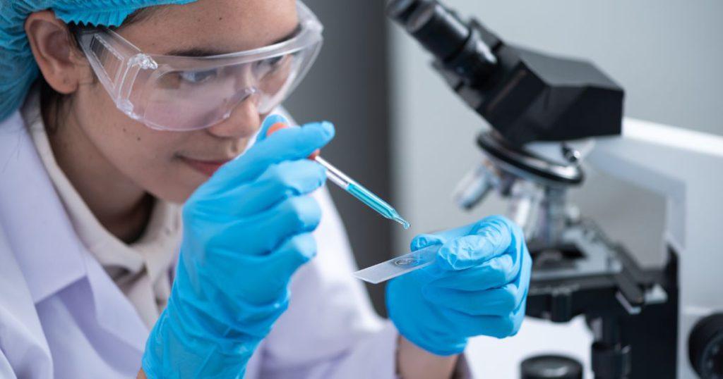
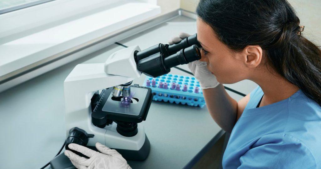
Welcome to the fascinating world of Clinical Microscopy, where we delve into the microscopic universe to identify bacteria, fungi, parasites, and viruses. This field of study is crucial for diagnosing diseases, understanding their causes, and developing effective treatments.
Please note, this is a work in progress, and we’re continually adding new content to enrich your understanding of this complex field.

Identification of Microorganisms
The first step in understanding any disease is identifying the culprit. Clinical microscopy allows us to identify various microorganisms, including:
- Microscopic Identification of Bacteria
- Microscopic Identification of Fungi
- Microscopic Identification of Parasites
- Microscopic Identification of Viruses
Each microorganism has unique characteristics that make it identifiable under the microscope, and understanding these traits is key to effective diagnosis and treatment.

Staining Techniques and Interpretation
Staining is a technique used to enhance contrast in microscopic images, making it easier to identify specific structures or organisms. Here are some of the staining techniques we’ll explore:
- Acid-fast Staining (Ziehl-Neelsen, Kinyoun)
- Fungal Staining (KOH, Calcofluor White)
- Gram Staining and Interpretation
- Immunohistochemistry and Immunofluorescence Staining
- Staining in Clinical Microscopy: Revealing the Invisible
- Giemsa Staining and Interpretation
- Hematoxylin and Eosin (H&E) Staining and Interpretation
- Wright Staining and Interpretation
Each staining technique has its unique purpose and interpretation, which we will delve into in each respective post.
Examination of Biopsies
Biopsies provide a snapshot of the disease in action. By examining tissue samples under the microscope, we can gain valuable insights into the disease process. Here are some of the biopsies we’ll examine:
- Microscopic Examination of Adenocarcinoma Biopsies
- Microscopic Examination of Basal Cell Carcinoma Biopsies
- Microscopic Examination of Bone Biopsies
Each biopsy examination provides a unique perspective on the disease, helping us understand its progression and impact on the body.
This post is part of my Clinical Microscopy category. Please check out index page on Clinical Microscopy
Further reading
Sean Schepers is a third-year Medical Technology student at Mahidol University with a passion for all things health and medicine. His journey into the world of medicine has led him to explore various fields. Sean's blog posts offer a unique perspective, combining his academic insights with personal experiences. When he's not studying or blogging, Sean enjoys keeping up with politics and planning his future career in medicine.
In addition to his studies, Sean serves as the chairman of the Rights, Liberties, and Welfare Committee, a role that reflects his commitment to advocacy and social justice. Beyond his academic pursuits, Sean offers tutoring services in English and Biology, further demonstrating his dedication to education and mentorship. His journey is one of continuous discovery, and he invites others to join him as he explores the dynamic and transformative world of medical technology.


